Athlete's heart 3D model design based on MRI data
Фотографии:

ˑ:
Assistant S.V. Strelkov1
Assistant A.L. Avtyushenko1
Professor, Dr.Sci. (Phys.-Math.) V.M. Ivanov1
Research scientist A.A. Solovyeva2
Dr.Med, Associate Professor N.V. Vasilyev3
1Peter the Great St. Petersburg Polytechnic University, Saint Petersburg
2The University Hospital Tübingen, Tübingen, Germany
3Boston Children's Hospital, Boston, USA
Keywords: MRI data processing, contouring, master-model design, augmented reality, 3d printing, visualization, cardiovascular diseases.
Introduction. Modern methods of medical visualization allow looking inside the human body without damaging it. It is especially relevant for athletes who are preparing for competitions and cannot interrupt their training program. A simple representation of three-dimensional graphical data of medical visualization methods still does not have a complete solution. The technology, described in this paper, is an effective solution which will enable the doctor to explain the anatomy and pathology of the patient's heart to the patient in a clearer way.
Objective of the research was to analyze and process MRI data of three different healthy volunteers to form a master-model of a healthy heart.
Methods and structure of the research. The main principle, used in the research, is based on creating a highly detailed master-model of the healthy heart with all internal structures and textures based on the analysis of various MRI data sets. This model will be used as the foundation for further creation of detailed models of patients' hearts with different deficiencies. Depending on the type of deficiencies, characteristic changes for certain pathologies are later made in the master-model.
The process of creating the master-model involves building up a basic model of the heart and adjusting its shape and anatomical properties according to the MRI data of several healthy hearts and subsequent formation of one final model. Primarily, an abstract model of the heart, based on anatomy atlases, is created. This model is then processed by 3D-artists jointly with doctors to obtain an accurate shape with all surface details and internal structures, such as valves and trabecularity, which cannot be fully reconstructed from the MRI data. Afterwards, the artists reconstruct the texture using photos or real hearts. As a result, a model of the heart is obtained, which will further be used as a basis. At this stage, the model is a basic shape which has all anatomical structures of heart but it still cannot be used as the master-model. The obtained model requires verification using the MRI data of a healthy person's heart.
At the next stage, MRI data sets of several healthy patients are obtained. Apart from standard data, the data set includes data of contours of the heart exterior as well as data of contours of the internal surface of ventricles and auricles. The contouring (hand-made delineation of heart's anatomical structures) is done by a professional doctor. This approach is one of the main ways for segmentation and determination of the contours of anatomical structures [2]. The contours are delineated at a certain stage of the cardiac cycle – during diastole in this case (Fig. 1).
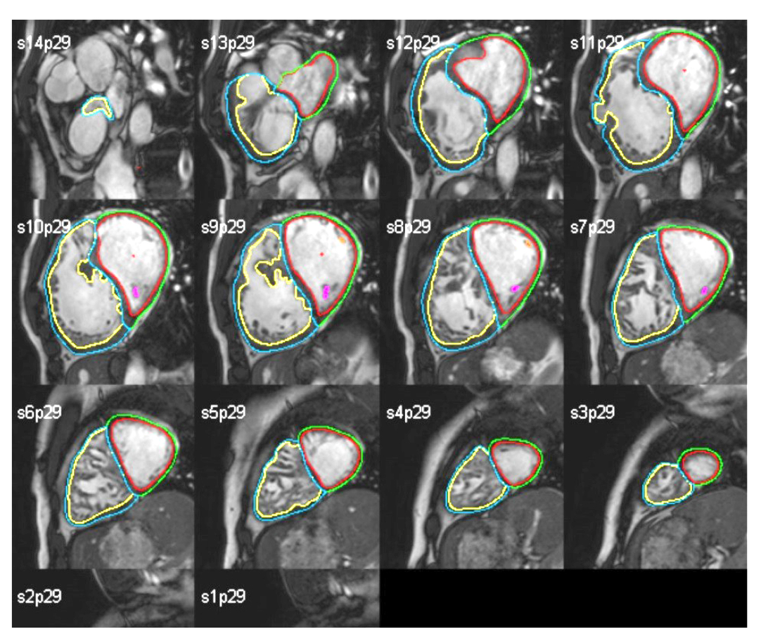
Fig.1.The contours data overlaying MRI
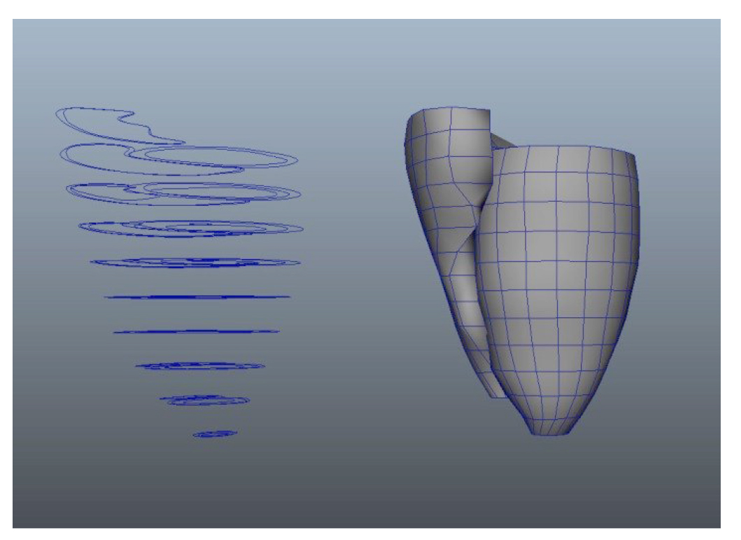
Fig. 2. Contours constructed in space and a 3D model based on them
On the basis of the contouring data analysis, a specialized software was developed which constructs curves in the space where the vertical axis distribution occurs on the basis of section distances in the MRI data set. According to these curves, the heart exterior and internal surfaces of ventricles are formed (Fig. 2). The MRI data often contain incomplete data – it has high noise and low spatial resolution. To enhance the images, different image noise removal algorithms [1] are applied, but in most cases this approach does not solve the problem. Thus, it is necessary to adopt the lacking details from a different source, which is the basic heart model created on the basis of the data from anatomy atlases. This approach requires comparison of the basic model with the model created on the basis of the contours. To solve this task, the basic model is uploaded to the model of contours and is aligned in horizontal and vertical planes with reference to the end points of ventricles. Then, the construction of a deformer grid is done along the perimeter of the basic model with the number of subdivisions equal to the number of layers of the MRI data and to the distance between them. Afterwards, the correlation of points of the deformer grid with points of the contours' cross-sections is done, whereas points which do not belong to the grid (the top and bottom of the model) are scaled proportionally by the average delta of grid point movement (Fig.3).
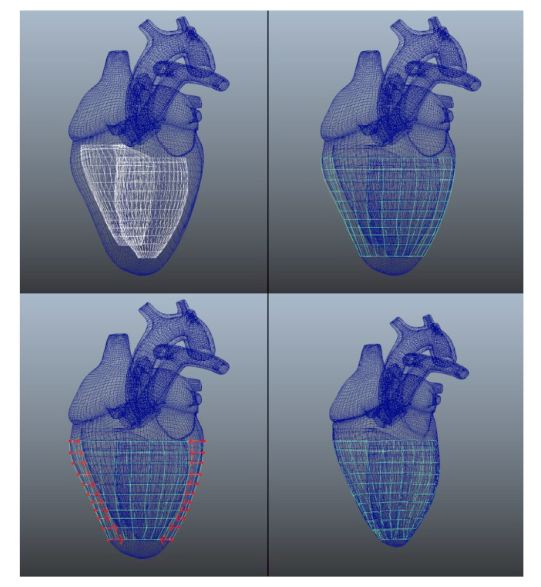
Fig. 3. Stages of the projection of the basic model upon the contours data
This approach to projection allows filling in the lacking fragments, which are not present in the MRI data, and at the same time saving the available information on external and internal contours of the heart and ventricles.
Then, to optimize the master-model, the data of three healthy patients were processed according to the algorithm described above (Fig.4). The result of averaging out of these models is the master-model shown in Fig. 5.
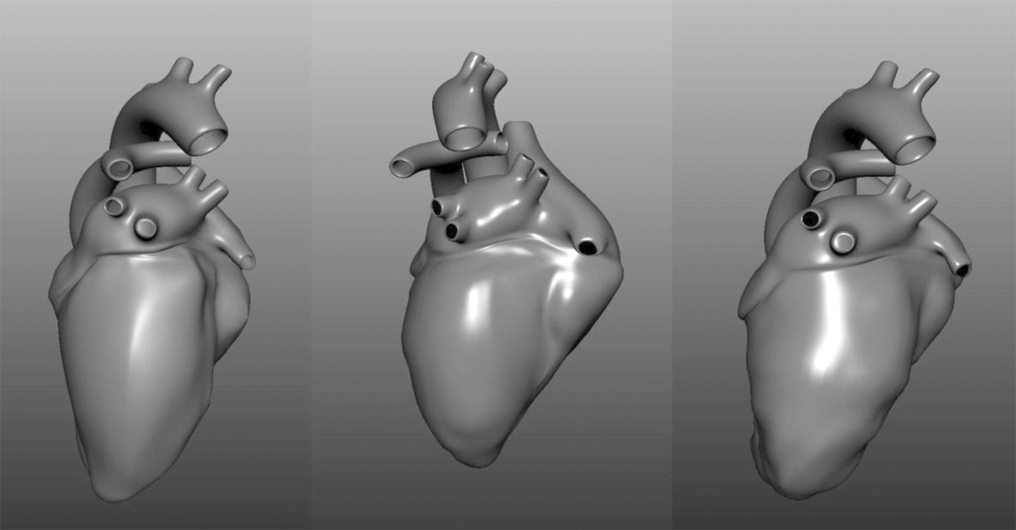
Fig. 4.
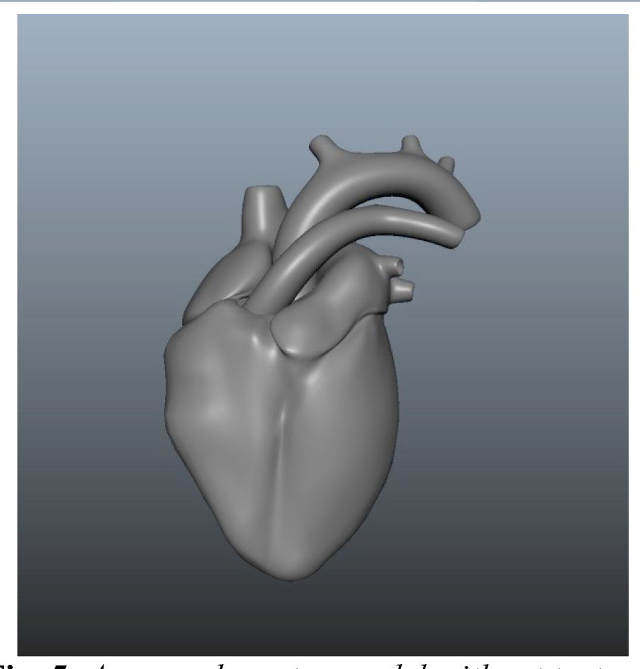
Fig. 5. Averaged master-model without textures
Results and discussion. As a result, this model is specified and the image of the heart of a certain patient is formed. For this purpose, the MRI data of this particular patient are used and a model of the patient's heart is constructed using the algorithm described above. In this case, an averaged model of the healthy heart is taken as a basis and not an abstract one, created according to anatomy atlases.
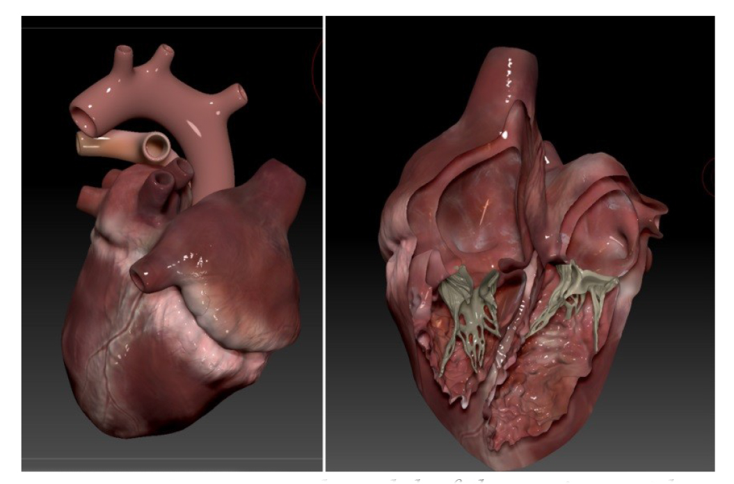
Fig. 6. Constructed model of the patient with internal structures and texture
Thus, a detailed model of the heart is obtained, which conforms to the heart shape of a particular patient and has the required detail and texture quality (Fig.6). It is obvious that the result obtained in this way is not a perfectly accurate 3D representation of the patient's heart. However, within the context of the set tasks, in which the doctor needs to explain the state of the damaged organ in terms of its anatomy, this solution is optimal. Besides, this algorithm can be automated.
Conclusion. In conclusion, it should be noted that the approach described above is successfully applied in design and visualization of a 3D model of heart. Besides, the developed approach with all developed related technologies can be easily adapted to visualize other organs of athletes. Thus, by using this solution, it is possible to display the state of the patient's organs and hereby to simplify the process of doctor-patient interaction.
References
- Paul Bao and Lei Zhang “Noise Reduction for Magnetic Resonance Images via Adaptive Multiscale Products Thresholding”, IEEE TRANSACTIONS ON MEDICAL IMAGING, vol. 22, no. 9, SEPTEMBER 2003, p. 1089.
- Caroline Petitjean and Jean-Nicolas Dacher “A review of segmentation methods in short axis cardiac MR images”, Medical Image Analysis, vol. 15, pp. 169-184.
- Suruchee V. Nandgaonkar, Prof. A.B. Raut “Comprehensive Study on Cloud Computing”, IJCSMC, vol. 3, no. 4, April 2014, pp.733–738
- Ronald T. Azuma “A Survey of Augmented Reality”, in press.
Corresponding author: fizkult@teoriya.ru
Abstract
This article describes a simple and effective approach to design of a highly detailed model of heart. The three-dimensional model design method is based on the use of ventricular contours data obtained by MRI study. This method allows building a model of an athlete's heart with all internal structures, which allows to simplify the process of visualization and assessment of cardiac structures before and after the load and preventing a possible pathology.


 Журнал "THEORY AND PRACTICE