Physiological features of shot technique of football players with musculoskeletal disorders
Фотографии:
ˑ:
Postgraduate student M.S. Nagornov2
Ph.D., Associate Professor K.V. Davlet'yarova2
Ph.D. A.A. Il'in3
Dr.Med., Professor L.V. Kapilevich1
1National Research Tomsk State University
2National Research Tomsk Polytechnic University
3Tomsk State University of Control Systems and Radioelectronics, Tomsk
Keywords: football, motor disorders, scoliosis, platypodia, training
Introduction
It is a common knowledge today that modern football is one of the most demanding sports that requires high individual physical and mental fitness from the players. Regular football sessions are known to help develop high agility, speed, motor coordination, motor responsiveness and spatial orientation in the players. Moreover, ball games on the whole and football in particular help foster team-work abilities and skills and cooperativeness in the players plus high self-control, determination, courage and excellent physical health [4].
As things now stand, however, the proportion of really healthy young people in the population is very low, but most of the children and adolescents experiencing different health disorders tend to stand against the situation and the relevant health restrictions in their socializing endeavours including sport activities [6]. This is the reason why many active footballers among university students are diagnosed with different musculoskeletal system pathologies including scoliosis, osteochondropathy, remissive osteochondrosis, platypodia etc. [1].
This situation requires give more attention to the effects of musculoskeletal system disorders on the bodily functions of an athlete to identify possible functional regularities in the body systems and, based on the study data obtained, offer the relevant adjustments to the standard football training systems.
The purpose of the study was to study the specific bioelectrical activity in the lower limb muscles and the relevant motor coordination sequences for the shots made using the inside of the foot by the footballers having musculoskeletal disorders including the II-III category scoliosis and the II-III degree platypodia.
Materials and methods
Subject for the study were the 1st to 3rd-year students (n=50) of 18 to 23 years of age playing for the National Research Tomsk Polytechnic University and Tomsk State University, each of them having football skills good enough to qualify for the first university team. We split up the volunteers equally into Experimental Group (n=25) and Control Group (n=25). Control Group was formed of the footballers free of any known musculoskeletal disorders, whilst the students having spinal (the II-III category scoliosis) and foot (II-III degree platypodia) pathologies were attributed to Experimental Group. The football training schedules and workloads were identical for both of the Groups. In the study tests, the footballers were requested to shoot motionless ball using the inside of the foot.
Method of computer stabilography was used to assess the player’s postural equilibrium and motor coordination during the test by means of “Stabilan 1” Computer Stabilographic Analyzer system. The athletes performed the shots using the inside of the foot standing on a platform of the Stabilographic Analyzer system, with the following performance indices being registered during the tests: Qx that means the dispersion (value of deviation) of the pressure center over the frontal plane, mm; Qy meaning the dispersion (value of deviation) of the pressure center over the sagittal plane, mm; mean speed of pressure center movement, mm/s; ellipse area, sq mm; Equilibrium Function Quality (EFQ), %; and mean linear speed, mm/s.
Muscle bioelectrical activity data during the shot were obtained using the Neuro-MVP-4 Computer Electroneuromyograph. Electrodes were placed as follows: Electrode #1 on the caput laterale of m. gastrocnemius; Electrode #2 on the caput mediale of m. gastrocnemius; Electrode #3 on the lower third of m. vastus lateralis; and Electrode #4 on the upper third of m. vastus lateralis.
Actual test data was presented in a mean ± error-of-mean format (М±m). Reliability of the group differences was estimated using the Mann-Whitney non-parametric test.
Results and discussion
The key postural equilibrium indices of the shots performed by Experimental Group football players (having musculoskeletal disorders) were found to have the following differences from that by Control Group (Table 1).
Table 1. Shots using the inside of the foot: stabilographic test data (X±m)
|
Indices |
Experimental Group |
Control Group |
|
Dispersion over the front plane, mm |
32,4±5,1* |
45,2±2,1 |
|
Dispersion over the sagittal plane, mm |
14,4±5,5* |
29,5±4,7 |
|
Pressure center mean movement speed, mm/s |
4,4±3,1* |
10,6±2,5 |
|
Area of ellipse, sq mm |
92,0±6,1* |
148,1±13,2 |
|
Equilibrium Function Quality (EFQ), % |
5907,3±1055,6* |
16296,5±3584,2 |
|
Mean linear speed, mm/s |
1031,6±124,7* |
2908,5±748,1 |
|
Equilibrium Function Quality (EFQ), % |
25,7±2,9* |
12,8±1,6 |
|
Dispersion over the front plane, mm |
92,6±6,5* |
149,3±13,3 |
*Group differences are reliable with p <0.05
Dispersion over the front plane for Experimental Group (32.4±5.1) was reliably lower (p < 0.05) than that for Control Group (45.2±2.1). Dispersion over the sagittal plane for Experimental Group players having musculoskeletal disorders (14.4±5.5) was reliably lower (p < 0.05) than that for healthy Control Group (29.5±4.7). Pressure center mean movement speed for the healthy Control Group players (10.6±2.5) was reliably higher (p < 0.05) than that for the Experimental Group players with the musculoskeletal disorders (4,4±3,1).
It is obvious from the above that the Experimental Group players showed some coordination disorders of detrimental effect on the movement linearity. This effect is due to the displacement of the body gravity center by the football players having musculoskeletal disorders. The center of pressure path is found to be distorted in every phase of the shot motor sequence. The players, however, tend to offset these distortions by adjustments that push down the few other performance indices including the dispersions over the frontal and sagittal planes, areas of the ellipse, mean linear speeds and the pressure center mean movement speeds [3, 5].
Present on Figure 1 hereunder are the lower limb muscle bioelectrical activity data for the shots performed using the inside of the foot by the Experimental Group vs. the Control Group players. The Experimental Group players having musculoskeletal disorders show reliably lower (p < 0.05) maximum amplitudes of the thigh muscles and the caput mediale of m. gastrocnemius activity data. However, the players having musculoskeletal disorders show higher bioelectrical activity data amplitudes for the caput laterale of m. gastrocnemius (1594.4 ± 121.1).
Therefore, the study data is indicative of the fact that the players having musculoskeletal disorders tend to perform shots by the inside of the foot using mostly shin muscles dominated by the caput laterale of m. gastrocnemius whilst the healthy football players perform the same shots using mostly the m. vastus lateralis.
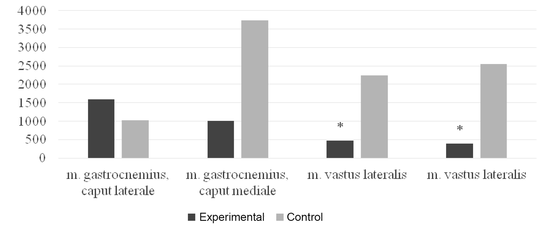
Lower limb muscle bioelectrical activity data for the shots performed using the inside of the foot:
Experimental Group (black) vs. Control Group (grey)
*Group differences are reliable with p <0.05
Conclusion
The players having musculoskeletal disorders show significant displacements of the body center of gravity compared to the healthy ones, and the specific motor coordination in the shooting sequence is distorted as a result. The players tend to compensate these dysfunctions by reducing the movement speed, but this compensatory mechanism results in the shot power falling too [2, 7].
The players try to offset the losses in the shot power by adjustments in the inter-muscular coordination pattern in the shooting lower limb. Most of the work is being taken over in the process by the shin muscles dominated by the caput laterale of m. gastrocnemius.
Relevant adjustments to the regular football training systems to make emphasis on the shin muscle workout and postural equilibrium exercises are recommended to help improve the shooting skills of the football players having musculoskeletal disorders including scoliosis and platypodia.
References
- Dubrovskiy, V.I. Patologicheskaya biomekhanika (Abnormal biomechanics) / V.I. Dubrovskiy, V.N. Fedorova // Biomekhanika: ucheb. dlya sredn. i vyssh. ucheb. zavedeniy (Biomechanics: textbook for sec. and higher. educ. institutions.). – Moscow: VLADOS – PRESS, 2003. – P. 591–628.
- Illarionova, A.V. Osobennosti vnutrimyshechnoy i mezhmyshechnoy koordinatsii pri dozirovanii usiliy v usloviyakh neustoychivogo ravnovesiya (Features of intramuscular and intermuscular coordination when dosing power at unstable equilibrium) / A.V. Illarionova, L.V. Kapilevich // Teoriya i praktika fiz. kul'tury. – 2014. – № 12. – P. 44–46.
- Kapilevich, L.V. Fiziologicheskoe obespechenie tochnosti i koordinatsii dvizheniy v usloviyakh neustoychivogo ravnovesiya i podvizhnoy tseli (Physiological support of accuracy and coordination in case of unstable equilibrium and moving target) / L.V. Kapilevich, F.A. Guzhov, Yu.P. Bredikhina, A.A. Il'in // Teoriya i praktika fiz. kul'tury. – 2014. – № 12. – P. 22–24.
- Koshel'skaya, E.V. Fiziologicheskie i biomekhanicheskie kharakteristiki tekhniki udarno-tselevykh deystviy futbolistov (Physiological and biomechanical characteristics of technique of targeted kick actions in football) / E.V. Koshel'skaya, L.V. Kapilevich, V.N. Bazhenov et al. // Byulleten' eksperimental'noy biologii i meditsiny. – 2012. – V. 153. – № 2. – P. 235–237.
- Skvortsov, D.V. Klinicheskiy analiz dvizheniya. Stabilometriya (Clinical analysis of movement. Stabilometry) / D.V. Skvortsov. – Moscow: Antidor, 2000. – 192 P.
- Shelkov, O.M. Mediko-biologicheskoe obespechenie paraolimpiyskikh vidov sporta. «Sportivnaya meditsina. Zdorov'e i fizicheskaya kul'tura. Sochi 2011» (Biomedical support of Paralympic sports. "Sports Medicine. Health and physical culture. Sochi 2011") / O.M. Shelkov, O.A. Churganov / Mater. II Vseros. (s mezhdunar. uchastiem) nauch.-prakt. konf. (Proc. of Rus. conf. (with intern. part.) June 16-18 2011 / Ed. by S.E. Pavlov. – Sochi, 2011. – P. 114–117.
- Geisser M.E. A meta-analytic review of surface electromyography among persons with low back pain and normal, healthy controls / M.E. Geisser, M. Ranavaya, A.J. Haig // Тhe Journal of Pain. – 2005. –N 6(11). – Р. 711–726.
Corresponding author: kapil@yandex.ru


 Журнал "THEORY AND PRACTICE