Strength display during gymnastic exercises
Фотографии:

ˑ:
Ph.D. D.V. Semenov
Associate professor, Ph.D. V.N. Shlyakhtov
A.A. Rumyantsev
Velikie Luki State Academy of Physical Culture and Sport, Velikie Luki
Keywords: strength qualities, gymnastic exercises, electromyographic activity, maximum voluntary muscle contraction.
Introduction. In sports practice among the methods of recording muscle strength in laboratory and natural conditions the most objective and convenient one is the electromyographic method comprising placement of surface electrodes on the skin of the subject and recording the electrical impulses generated by the muscles of an athlete while performing movements. Electromyogram (EMG) recording provides a relatively accurate idea of the order of muscle activation and indirectly reflects the strength of the main working muscles ensuring the performance of a specific sports motor action [1].
The purpose of the research was to determine the characteristics of leg muscles activity of gymnasts when doing a take-off to standing somersault, the amplitude of the electrical activity of the muscles, the order of their activation, duration and coherence of their work, as well as in comparison of EMG when performing a standing somersault to EMG recorded during maximum voluntary muscle contractions (MVC).
Materials and methods. Six gymnasts of the I category, candidates for master and masters of sport 15 to 21 years of age were examined. Each of them made 5 attempts of standing somersault in standard conditions (with a standard rest interval) followed by several attempts of more simple motor actions (vertical jump, series of jump-offs).
MVC of the lower leg and thigh muscles was recorded by means of the complex «Biodex Multi-Joint System Рго-3» (USA, 2006). Subjects performed ankle flexion and knee extension. When performing the plantar flexion of the foot the athlete was in an armchair, his right lower limb was fixed at an angle of 130° in the knee joint and 80° in the ankle joint. On a signal the athlete performed isometric contractions of his lower leg muscles. When testing the concentric contraction in the lower leg muscles the successively set load was 40%, 50%, 70%, 90% and 100% of the isometric maximum recorded. At the same time the subject moved the lever of the dynamometer by straightening his leg. Testing of isometric and concentric contractions in the thigh muscles was performed according to the same protocol with the athlete positioned in the armchair with the knee joint angle of 90°. Two movement attempts were performed in each case.
The amount of muscle effort was determined by the amplitude of their electrical activity recorded directly during the performance of the above movements using a 16-channel telemetric electromyograph "ME6000". Electromyogram for 6 pairs of lower limb muscles was recorded: tibialis anterior muscle (m. Tibialis anterior), soleus muscle (m. Soleus), gastrocnemius (m. Gastrocnemius) medial (medialis) and vastus lateralis (lateralis) as well as rectus femoris (m. Rectus femoris) and vastus external muscle (m. Vastus lateralis).
To determine the phase structure of the movement a kinematic analysis was conducted using the Qualisys complex. The data obtained in the course of the kinematic analysis of a standing somersault and corresponding to body positions of a gymnast when performing a take-off were used to determine the values of joint angles in order to simulate body positions of athletes while recording MVC using the «Biodex» complex.
Results and discussion. The following major phases were singled out while analyzing the biomechanical structure of a standing somersault: the preparatory phase – a movement from the starting position of standing on straight legs downward (squatting) (Figure 1 – А), the start of which is defined as downward movement of the trochanteric fossa; the main phase – legs extension and flying upwards taking a tuck position or jumping characterized by an upward movement of the trochanteric fossa (Figure 1 – B); the final phase – opening of the body and landing determined by a downward movement of the trochanteric fossa. The main phase in turn is divided into two stages – a take-off and flying upwards, bordering at the moment when feet’s contact with the supporting surface comes to an end (Figure 1 – C), and the start of the flight is associated with the start of the movement of the “final” point on the feet.
The average values of the joint angles by phase were as follows: at the beginning of the movement – hip - 165°, knee - 171°, ankle - 91°; in the early phase of the take-off (end of squatting) – hip - 96°, knee - 96°, ankle - 76°. At the end of the take-off – hip - 165°, knee - 134°, ankle - 114°.
In the course of the biomechanical analysis it was found that the duration of the take-off phase when performing a standing somersault is 0.2 seconds.
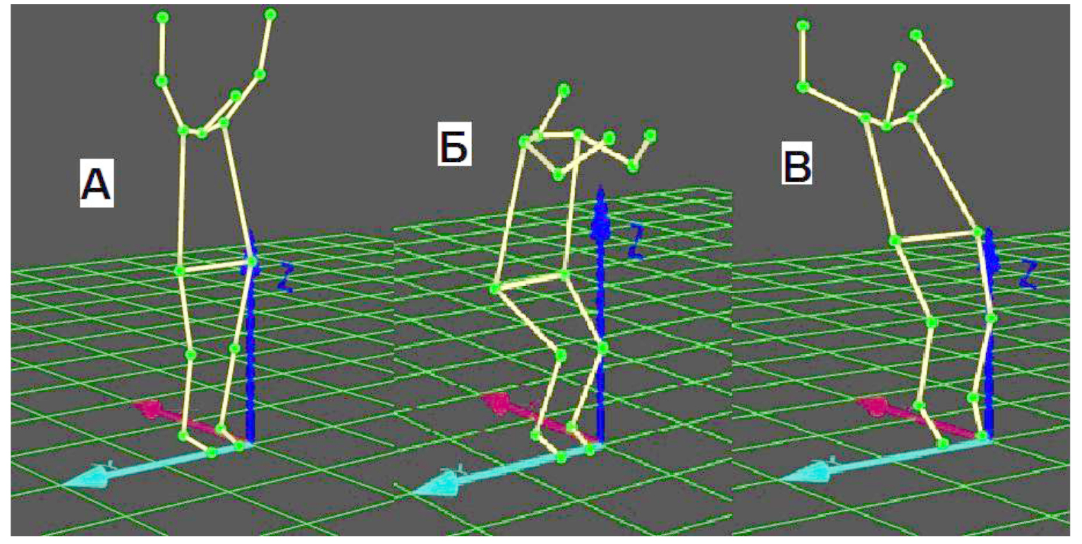
Figure 1. Take-off phases when performing standing somersault.
Proceeding from the EMG analysis, the level of electrical activity averaged at 135 microvolt during the preparatory phase of the movement – the squatting – associated with the eccentric contraction in the leg muscles. There is a sequential activation of the anterior tibial muscle followed by that of both the soleus and gastrocnemius muscles and then of the rectus and vastus external muscles during this phase. In the main phase (the take-off) there are almost simultaneous concentric contractions in the extensor muscles of the lower leg and thigh that reach the EMG values of over 300 microvolt. Activity of the lower leg muscles does not stop until the legs are fully extended and the contact with the supporting surface comes to an end, the thigh muscles exhibit the peak of their activity at the beginning of the take-off, and the duration of their activity does not exceed 0.1 second (Figure 2).
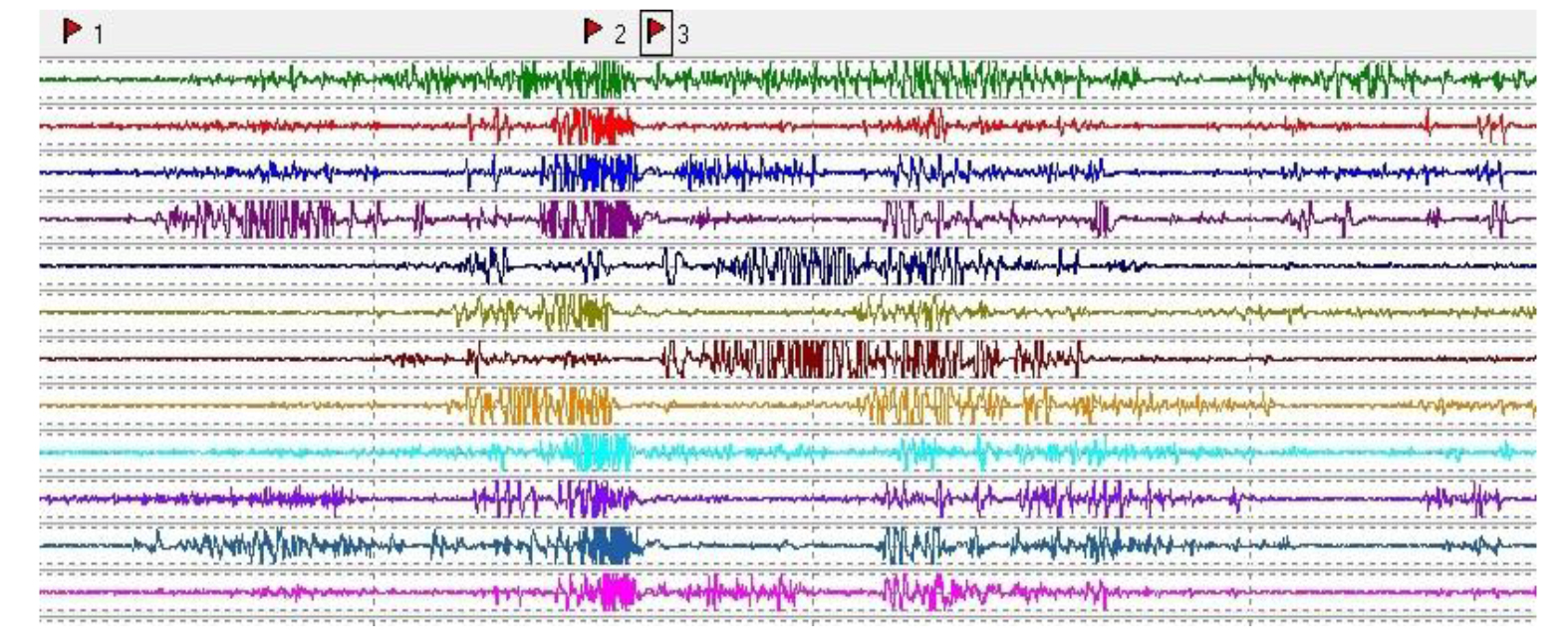
Figure 2. Electromyogram of the lower limbs muscles activity during gymnast's performance of standing somersault. Markers: 1 – the start of the preparatory phase (squatting), 2 – the start of the main phase (take-off), 3 – the moment of separation of feet from the supporting surface
It has been found that the amplitudes of the electromyogram of the lower leg and external vastus muscles registered during the take-off to standing somersault were higher than the values of the electromyogram of the muscles during maximal voluntary isometric and concentric contractions using the complex «Biodex», with the value of the electromyogram during concentric contractions being higher than during isometric ones (Figures 3, 4).
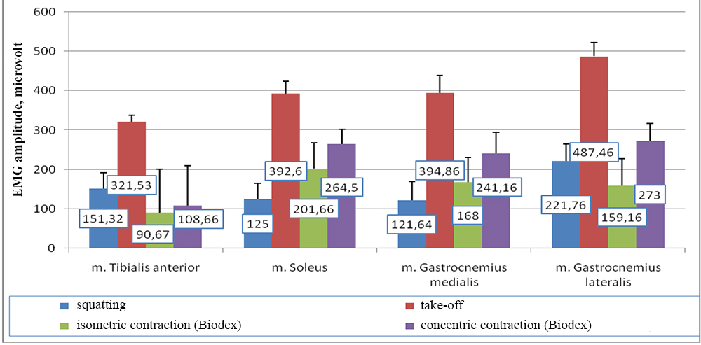
Figure 3. Comparison of average electrical activity in the lower leg muscles when performing standing somersault and plantar flexion of the foot using the “Biodex” complex in isometric and concentric contraction types
At the same time, one can observe in Figure 4 that in case of the rectus femoris we managed to record higher electromyogram values in isometric (314.16 microvolt) and concentric (304.5 microvolt) contractions compared with the electromyogram recorded when performing a standing somersault (123.36 microvolt).
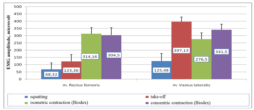
Figure 4. Comparison of the average electrical activity of the hip muscles when performing a standing somersault and knee extension using Biodex in isometric and concentric contraction types
The electromyographic analysis registered when the gymnasts were performing maximal voluntary muscular contractions of different types had not revealed any significant differences between the maximal concentric efforts and those 50% of the maximum in the lower leg and external vastus muscles regarding the degree of their electrical activation (Figure 5). The electrical activity of the rectus femoris during maximal isometric contraction is more intensive than during concentric one (Figure 5).
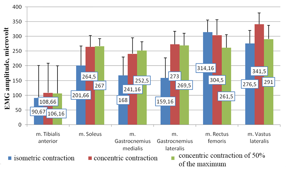
Figure 5. Comparison of the average electrical activity of the lower leg and hip muscles when performing voluntary muscle contractions of various types using “Biodex”
The results of statistical processing of the data obtained in the course of the research indicate high EMG variability, but enable us to trace certain regularities manifested in higher EMG values of muscles during concentric contraction, compared with the eccentric and isometric ones (Table 1).
Table 1. EMG (microvolt) of the lower limb muscles of gymnasts when performing standing somersault and during maximum voluntary contraction of examined muscles using “Biodex” complex
|
Standing somersault |
Statistical indicators |
Studied muscles |
|||||
|
m. Tibialis anterior |
m. Soleus |
m. Gastrocnemius medialis |
m. Gastrocnemius lateralis |
m. Rectus femoris |
m. Vastus lateralis |
||
|
|
Preparatory phase – squatting (eccentric contraction) |
||||||
|
М |
151.32 |
125 |
121.64 |
221.76 |
68.32 |
125.48 |
|
|
|
Take-off (concentric contraction) |
||||||
|
М |
321.53 |
392.6 |
394.86 |
487.46 |
123.36 |
397.13 |
|
|
Biodex |
|
Plantar flexion of the foot (isometric contraction) |
|||||
|
М |
90.67 |
201.66 |
168 |
159.16 |
21.66 |
75.83 |
|
|
|
Plantar flexion of the foot (concentric contraction) |
||||||
|
М |
108.66 |
264.50 |
241.16 |
273.00 |
33.16 |
112.83 |
|
|
|
Knee extension (isometric contraction) |
||||||
|
М |
86.50 |
97.16 |
45.16 |
61.83 |
314.16 |
276.50 |
|
|
|
Knee extension (concentric contraction) |
||||||
|
М |
130.66 |
60.50 |
30.00 |
33.50 |
304.50 |
341.50 |
|
Note: EMG values of the studied muscles are highlighted in bold.
As seen from the data shown in Table 1 (highlighted cells), while performing a standing somersault in the take-off phase five out of six studied muscles develop more intensive efforts than those when performing maximum voluntary muscle contractions using the Biodex” complex, as shown by a higher EMG. The EMG amplitude of the muscles is higher in case of isometric contractions than in case of eccentric ones, but is lower than that of concentric contractions, which is also supported by research of other authors [2, 4]. A similar pattern was also observed in our research of electrical activity in muscles when doing back flip in acrobatics [3]. All the studied gymnasts had EMG amplitudes of the gastrocnemius muscle higher when doing back flip than when performing maximum voluntary muscle contractions using the “Biodex” complex.
Moreover, in the course of the research there was some internal asymmetry of muscle activity manifested in the higher EMG of the left hip muscles compared with the muscles of the right one and higher EMG values of the right lower leg muscles compared with those of the left lower leg which in general did not show in the external picture of the movement.
Conclusion. Recording of a higher EMG amplitude when performing a real movement can be due to the characteristics of the implementation of a complex coordinated motor skill that involves mobilization and coordinated work of all the muscle groups involved in the movement. This fact can also be explained by that when systematic long-lasting training takes place, a motor command probably develops in the cortex of a gymnast that leads to activation of a larger number of high-threshold motor units and, consequently, to the development of significant muscular efforts.
The findings of the study can be used when selecting techniques of strength training depending on the types of contraction of core muscles, ensuring the performance of motor actions.
References
- Gorodnichev, R.M. Sportivnaya elektronejromiografiya (Sport electroneuromyography) / R.M. Gorodnichev. – Velikie Luki, 2005. – 300 P.
- Samsonova, A.V. Kharakteristika summarnoy elektricheskoy aktivnosti myshts pri vypolnenii silovykh uprazhneniy (Characteristics of total electrical activity of muscles during weight training) / A.V. Samsonova // Visnik Chernigivs'kogo derzhavnogo pedagogichnogo universitetu. Vipusk 81. Seriya: Pedagogichni nauki. Fizichne vikhovannya ta sport. – Chernigov, 2010. – P. 427–431.
- Shlyakhtov, V.N. Sravnitel'ny analiz myshechnykh usiliy pri vypolnenii perevorota nazad v akrobatike i proizvol'nykh dvizheniy (Comparative analysis of muscular efforts when doing back flip in acrobatics and voluntary movements) / V.N. Shlyakhtov // Teoriya i praktika fizicheskoy kul'tury. – 2013. – № 11. – P. 79.
- Gibala, M.J. Changes in human skeletal muscle ultrastructure and force production after acute resistance exercise / M.J. Gibala, J.D. MacDougall, M.A. Tarnopolsky, W.T. Stauber, A. Elorriaga // Journal of Applied Physiology, 1995. – V. 78. – P. 702–708.
Corresponding author: gorodnichev@vlgafc.ru


 Журнал "THEORY AND PRACTICE