Neuromuscular Apparatus of Female Basketball Players in View of Their Age Features
Фотографии:

ˑ:
A.V. Sysoev, Ph.D., pro-rector for sports
I.E. Popova, associate professor, Ph.D.
Voronezh State Institute of Physical Culture, Voronezh
Key words: neuromuscular transmission, excitability of motor neurons, functional features.
Introduction. The specificity of the motor mode of people involved in sports influences functioning of all body systems, including neuromotor apparatus. Regular sports occupations contribute to very accurate organization of regulation of performance of a motor act, fully complying with necessary motor tasks. Here, the study of the peculiarities of functioning of the neuromuscular apparatus under the influence of specific sports activity contributes to a more detailed analysis of the mechanisms of development of adaptive processes in athletes' neuromotor system as well as their possible restructuring due to some factors associated with the implementation of physical load of various volumes [9].
In the process of age-related development human neuromuscular apparatus undergoes structural and functional changes at neural, muscular, hormonal and biochemical levels. Since an athlete cannot be trained without the formed motor function, more sophisticated technologies are required, that take into account not only the specificity of sports activity, but also age and initial level of athletes' motor fitness [8, 12, 13]. The level of the motor component of functional fitness can be objectively estimated by means of electromyography [5]. It is also known that the enhancement of sports skills promotes an improvement of regulation mechanisms, body's functionalities and motor coordination. Thus, the purpose of the research was to study the functional features of the neuromuscular apparatus of female basketball players of different age groups and sports qualifications acquired during the long-term training process.
Materials and methods. The subjects of the research were female basketball players divided into 3 groups according to age. The first one consisted of the athletes aged 13-14 (n = 10), the second one - of 15-16 year olds (n = 10), and the third one - of 19-30 year olds (n = 10). The obtained data were compared to the standards provided in the cited literature [1, 3, 4, 6, 7, 11].
The bioelectrical activity of muscles was evaluated using the neuromyoanalyzer NMA-4-01 "Neuromyan". The basic study method was stimulation myography of the following nerves: Medianus, Ulnaris, Musculocutanius, and Tibialis. Herewith, the M-response of the Abductor pollicis brevis, Abductor digiti minimi, Abductor hallucis and Biceps brachii muscles was recorded.
In order to record the motor response of the muscles (M-response), the surface electrodes were used. The active electrode was placed at the muscle motor point, the reference one - at the tendon area of that muscle. The grounding electrode was applied between the lead and the stimulating electrodes. The stimulating bipolar electrode was placed over the projection of the nerve which innervated the given muscle. During the research on the nerve conduction, the supramaximal stimuli were used.
To evaluate the conductivity of the segmental arch, including the sensory and motor fibers outside the spinal cord and its intraspinal area, and also the motor neuron excitability, H-reflex of the medial head of the Gastrocnemius muscle was registered, and the reflex was provoked by a commonly used stimulation of the Tibialis nerve by means of the unipolar electrode, and with that, the active electrode was placed in the popliteal fossa [6].
The obtained data were processed by the conventional methods of the analysis of variance, performing the validation of different empirical samplings using the Student's criterion (t-criterion).
Results and discussion. To evaluate the total muscle electrical potential in response to a one-time electric stimulation of the motor fibers of the mixed peripheral nerve, the M-response of the upper and lower limb muscles of the female basketball players from different age groups was recorded. There was noted a statistically-valid reduction of the latency value, as well as of the residual latency (RL) of the M-response of the female athletes from Group 3, as compared to the ones from Groups 1 and 2, while stimulating the Medianus, Ulnaris, Musculocutanius and Tibialis nerves (Figures 1-4). The obtained data are indicative of the increase of the maximal conduction of excitation in the nerve fibers of the adult female basketball players having maximum sports experience, which enables better development of speed strength.
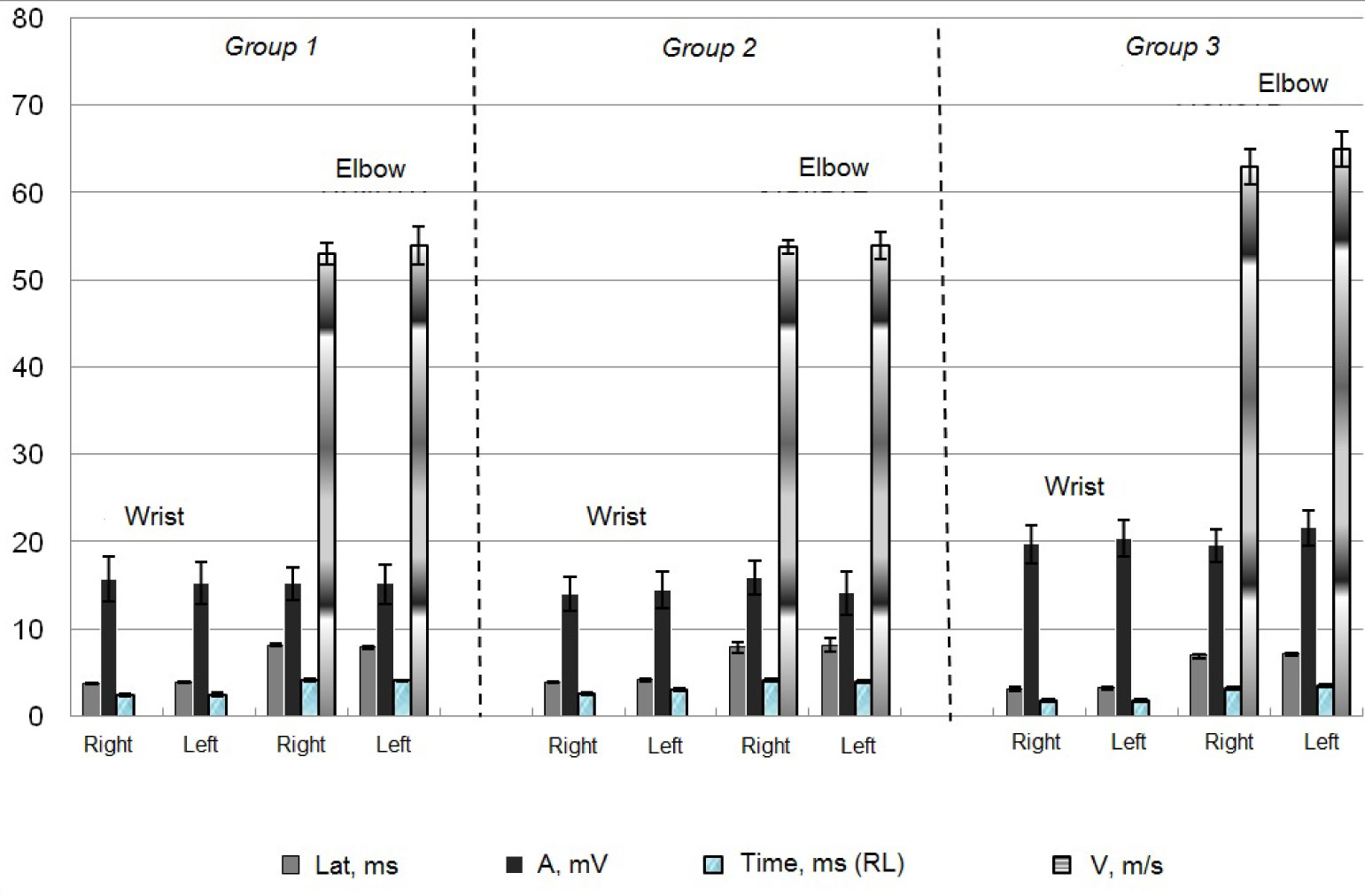
Figure 1. Characteristics of M-response during stimulation of Medianus nerve of female basketball players from different age groups.
The increase of the maximal conduction of excitation in the motor fibers of the upper and lower limbs of the female basketball players from Group 3 indicates an increase of their lability, which determines not only the shortest time needed for the excitation to appear, but also the time needed for the excitation to tone down and for the tissue to restore and produce the succeeding excitation pulses [1].
It is also known that the latent period of the motor reaction comes as an indicator of the fast muscle contraction speed [5]. Consequently, regular basketball occupations enable quality improvement of the athletes’ neuromuscular apparatus, which corresponds to the literature data [14]. Herewith, the cornerstone of the speed-strength movement is the ability of the muscles to stretch with momentary and significant intensity and then to contract fast and powerfully. Fast muscle contraction speed is one of the most important factors which determine the functional state of the muscular system of the athletes who specialize in speed-and-strength sports [10].

Figure 2. Characteristics of M-response during stimulation of Ulnaris nerve of female basketball players from different age groups.
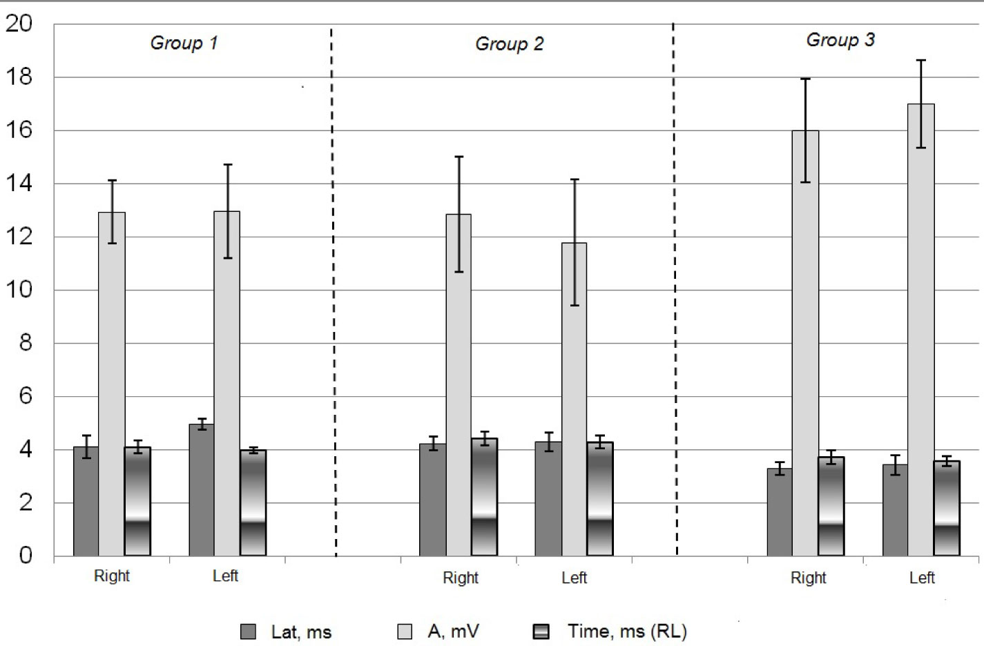
Figure 3. Characteristics of M-response during stimulation of Musculocutanius nerve of female basketball players from different age groups.
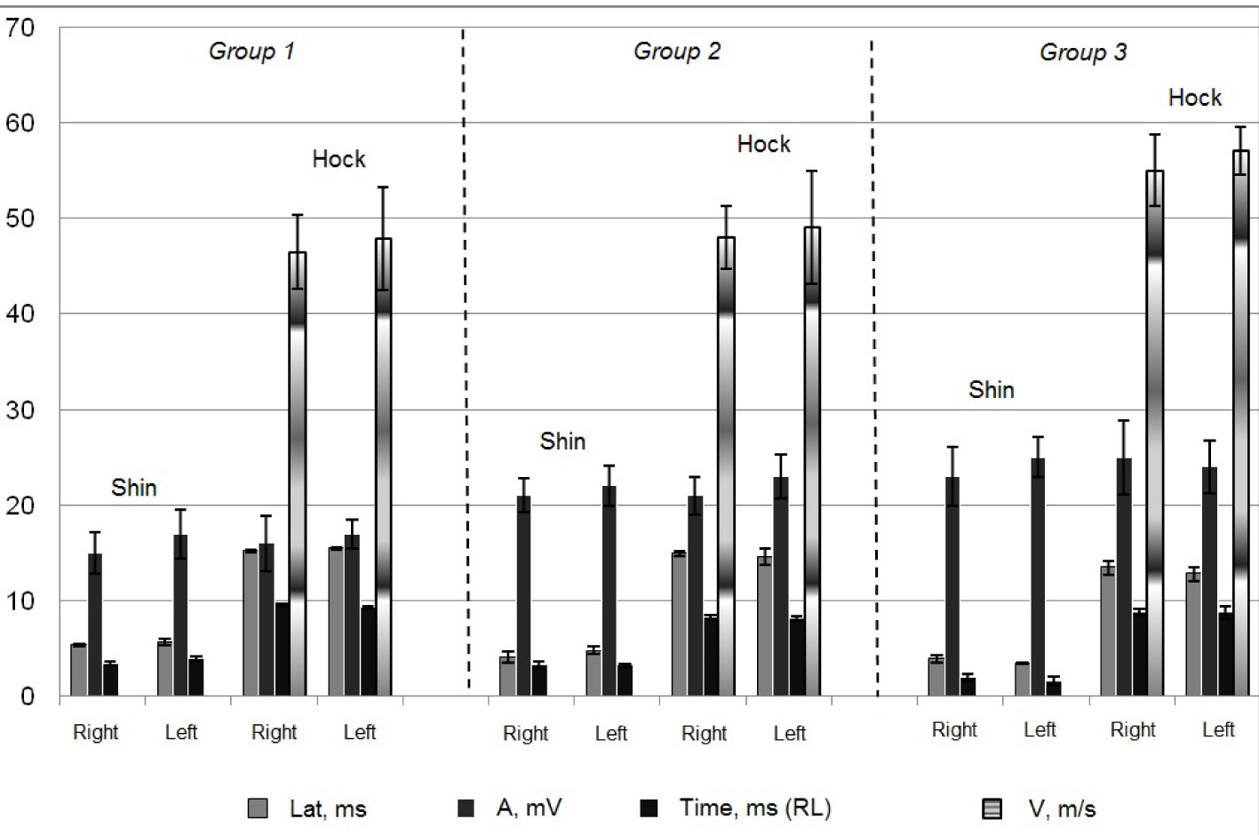
Figure 4. Characteristics of M-response during stimulation of Tibialis nerve of female basketball players from different age groups.
During the research on the M-response amplitude of the upper and lower limb muscles, its highest value was recorded among the more experienced female basketball players from Group 3 than that among the younger female athletes (Figures 1-3). According to the obtained data, the extension of time for basketball training and growing of female athletes contributed to the increase in quantity and synchronism of the involved muscles of the upper and lower limbs, which enabled the momentary force to develop at its maximum, and that will characterize the maximum dynamical force and, consequently, will help to improve sports results. The increase of the amplitude among the female athletes from Group 3 may also be conditioned by the expansion of the cross-section area of the muscle fibers as a result of regular training sessions.
Since by the age of 12-15 the muscle spindles stop growing, and the muscle tissue structure becomes the same as the one of adults aged 20-30 [1, 17], the detected distinctions in the functioning of the neuromuscular apparatus of the adult female basketball players are most probably conditioned by their long-term basketball occupations as opposed to the younger female athletes. Thus, the number and the development level of certain muscle spindles of different muscles are not identical, and their structural complexity depends on the amplitude of movement and force of muscle contraction, which is related to the muscle coordination [12]. Its volume is higher among more experienced female athletes.
The more qualified adult female basketball players were also determined to have the maximum speed of the nerve impulse conduction through the nerves under study (Figures 1-3). Most probably, that is conditioned by the long-term basketball occupations of the female athletes from Group 3, as by the age of 13 sensory and motor receptors of the muscular system reach their full growth and get similar to the ones of adults [1].
The synchronous reflexive response of the motor unit of the medial Gastrocnemius muscle to the electric stimulation of the sensory fibers of the Tibialis nerve was evaluated using the H-reflex method. The statistically significant differences of such characteristics of H-reflex as АMP Н/М and Lat are lacking (Table). The obtained data indicate a similar relative share of the reflexively excited motoneurons out of all medial Gastrocnemius muscle neurons.
Table. Characteristics of H-reflex of medial Gastrocnemius muscle of female basketball players from different age groups
|
Parameter |
Group 1 |
Group 2 |
Group 3 |
|
|
Н/М, % |
Left |
17,6±2,7 |
19,1±2,9 |
21,3±1,9 |
|
Right |
21,0±1,9 |
22,4±2,3 |
18,7±3,1 |
|
|
Lat, ms |
Left |
31,6±2,1 |
29,6±2,1 |
32,2±2,7 |
|
Right |
32,8±1,7 |
30,5±2,7 |
31,7±2,3 |
|
However, it has been established that by the age of 18-27 the H-reflex amplitude reduces [16]. Perhaps, the identified indistinction in the reflex excitability of α-motoneurons of female basketball players from different age groups is conditioned by the influence of the long-term basketball occupations of the adult athletes, whose level of sports qualification is higher and whose speed-strength development and formation and maintenance of the comprehensive coordination motor skills are better than those of the female athletes from Groups 1 and 2. The current hypothesis was confirmed by the literature data, according to which the excitation of the α-motoneurons is higher among the athletes with higher sports qualification [2,5, 15].
The registered mean-group latency values of the H-reflex do not differ significantly within different age groups and are within the normal limits for healthy people [3]. The obtained data indicate that the female basketball players of different ages have a similar degree of myelination and diameter of the sensory and motor fibers.
The fact in evidence is stipulated by the speed of the nerve impulse conduction during the fiber growth period, as well as by the specificity of the sports activity.
Conclusion. Hence, the analysis of the obtained data has revealed that long-term basketball occupations contribute to the increase of the maximum conduction of excitation in nerve fibers, the number and synchrony of active motor units of muscles of upper and lower limbs. The identified physiological features of the neuromuscular apparatus can serve as objective indicators for the operational control of the training process of young female athletes, where the stipulated qualities should be developed to improve the efficiency of athletes' playing actions in the early phases of basketball occupations. While training young female basketball players, it is necessary to use the exercises that help increase the share of the reflexively excited motoneurons out of all medial Gastrocnemius muscle neurons, and also the degree of myelination of the Tiabilis nerve, which enables to develop and improve the comprehensive coordination motor skills.
References
- Antonova, V.A. Anatomy and Physiology / V.A. Antonova. – Moscow: Vysshee obrazovanie. – 2006. – 192 P. (In Russian)
- Arifulin, A.N. Functional characteristics of neuromotor apparatus of lower limbs in young athletes of different specializations: Ph.D. thesis / A.N. Arifulin. - Vladimir, 2005. – 128 P. (In Russian)
- Badalyan, L.O. Clinical electroneuromyography / L.O. Badalyan, L.A. Skvortsov. – Moscow: Meditsina, 1986. – 368 P. (In Russian)
- Bezrukikh, M.M. Age physiology (Physiology of Child’s Development) / M.M. Bezrukikh, V.D. Son’kin, D.A. Farber. – Moscow: Academia, 2003. – 416 P. (In Russian)
- Gorodnichev, R.M. Sport electroneuromyography / R.M. Gorodnichev. – Velikie Luki: VSAPhCS, 2005. – 227 P. (In Russian)
- Komantsev, V.N. Methodological bases of clinical electroneuromyography / V.N. Komantsev, V.A. Zabolotnykh. – St.Petersburg, 2001. – 350 P. (In Russian)
- Kotz, Ya.M. Sport physiology / Ya.M. Kotz. – Moscow: Fizkultura i sport, 1986. – 135 P. (In Russian)
- Lanskaya, O.V. Age features of change in motor reflexes of human lower limb muscles / O.V. Lanskaya // Novye issledovaniya. – 2011. – № 1. – P. 15-21. (In Russian)
- Lanskaya, O.V. The study of bilateral monosynaptic reflexes muscles of upper and lower limbs in cyclic and game sports representatives / O.V. Lanskaya, E.Ya. Andriyanova // Novye issledovaniya. – 2012. – № 4. – V. 33. – P. 5-12. (In Russian)
- Makarova, G.A. Methodological principles of analysis and evaluation of physiological criteria of athletes’ functional state / G.A. Makarova, M.L. Igel’nik, V.V. Besschastnaya // Teoriya i praktika fizicheskoy kultury. – 2007. – № 10. – P. 49-52. (In Russian)
- Nikolaev, S.G. Workshop on clinical electroneuromyography / S.G. Nikolaev. - Ivanovo, 2003. – 264 P. (In Russian)
- Pryanishnikova, O.A. Sport electroneuromyography / O.A. Pryanishnikova, R.M. Gorodnichev, L.R. Gorodnicheva, A.V. Tkachenko // Teoriya i praktika fizicheskoy kultury. – 2005. – № 9. – P. 392. (In Russian)
- Sonkin, V.D. Development of muscular and energy working capacity in ontogenesis / V.D. Sonkin, R.V. Tambovtseva. – Moscow: LIBROKOM, 2011. – 368 P. (In Russian)
- Ter-Hovhannisyan, I.A. Training athletes: a modern view / I.A. Ter-Hovhannisyan. – Moscow: Terra-Sport, 2000. – 128 P. (In Russian)
- Fomin, R.N. Presynaptic inhibition of a-motor neurons of the spinal cord in athletes adapted to diverse motor activity / R.N. Fomin, D.K. Fomina // Teoriya i praktika fizicheskoy kultury. – № 9. – 2005. – P. 12-16. (In Russian)
- Chelnokov, A.A. Functional features of reflex and motor responses of skeletal muscles at rest in people of different ages / A.A. Chelnokov // Novye issledovaniya. – 2012. – № 4. – V. 33. – P. 13-22. (In Russian)
- Kido, A. Spinal excitation and inhibition decrease as humans age / A. Kido, N. Tanaka, R.B. Stein // Canadian Journal of Physiology and Pharmacology. – 2004. – V. 82. – № 4. -– Р. 238-248.
Corresponding author: delta8080@mail.ru


 Журнал "THEORY AND PRACTICE